Imaging of shoulder instability
Review of the article: Baudi P, Rebuzzi M, Matino G, Catani F. Imaging of the Unstable Shoulder [1].
Introduction
Shoulder instability is a fairly common condition in which the connective tissues around the glenohumeral joint, the ligaments and the joint capsule, are lax, so the bones forming the joint move through an excessive range. An unstable shoulder can arise for various reasons and present with many different symptoms, from an acute dislocation of the glenohumeral joint to chronic instability after repeated dislocations. A dislocation can cause different types of injury: to the ligamentous complex and/or to the articular parts of the shoulder girdle bones, on one or both sides of the joint. The diagnosis can often be made from the history and clinical signs, but imaging findings are decisive in choosing further treatment. Magnetic resonance imaging (MRI) with a field strength of at least 1.5 Tesla is the most widely used imaging tool for soft-tissue lesions of the shoulder (Fig. 1), although it may not reliably show some injuries, for example SLAP lesions (Superior Labrum Anterior to Posterior), tears of the superior labrum at the attachment of the long head of biceps. Magnetic resonance arthrography (MRA), which shows this injury better than standard MRI, is therefore used as well. In acute cases MRA is not needed, because the effusion and haemarthrosis act as a contrast medium.
When bone defects are present, CT should be performed. 3D CT images (Fig. 2) allow an objective assessment of the nature and extent of glenoid damage, and a 3D CT reconstruction with an en face view of the humeral head is the gold standard for assessing a Hill-Sachs lesion (a cortical depression in the posterolateral humeral head).
Careful selection and interpretation of imaging studies are therefore essential to detect, characterise and quantify the lesions. In the unstable shoulder, imaging is central to choosing the treatment method, planning surgery and preventing recurrence; patient factors such as individual biological and anatomical features and lifestyle (level of physical activity) should also be taken into account.
The rest of the article deals with anterior instability only; imaging of posterior and multidirectional instability is covered in separate chapters of the same monograph.
More relevant information is available via Diagnosis and treatment of posterior shoulder instability based on the ABC classification.
Labrum, ligament and capsule injuries: detection and assessment
MRI has always been considered the gold standard for assessing soft-tissue injury. MRA in particular showed high sensitivity (86–91%) and high specificity (86–98%) for the capsuloligamentous complex and cartilaginous structures. MRI is less sensitive and specific (44–100% and 66–95%, respectively) for detecting and assessing labral injuries. MRI is the gold standard for examining the rotator cuff, and MRA for partial-thickness cuff tears. MRA, however, is more expensive, takes longer, is more invasive and exposes the patient to ionising radiation when performed under fluoroscopic guidance.
Labral lesions are described simply by their position on the “glenoid clock face”. Several terms are used for anteroinferior labral injuries, based on their pathoanatomy:
- Bankart lesion: complete detachment of the labroligamentous complex from the glenoid with disrupted periosteum; it is the most common lesion after traumatic anterior dislocation;
- Perthes lesion: non-displaced tear of the anteroinferior labrum; the intact periosteum is lifted off the scapular neck, and the inferior glenohumeral ligament complex remains attached to it;
- ALPSA (anterior labroligamentous periosteal sleeve avulsion): avulsed inferior glenohumeral labroligamentous complex displaced medially along the glenoid neck, with intact periosteum;
- GLAD (glenolabral articular disruption): superficial tear of the anteroinferior labrum with injury to the adjacent articular cartilage;
- ALIPSA: avulsion of the anterior band of the inferior glenohumeral ligament from the glenoid, with an intact labrum;
- HAGL (humeral avulsion of the glenohumeral ligament): avulsion of the inferior glenohumeral ligament from its humeral insertion;
- BHAGL (bony humeral avulsion of the glenohumeral ligament): as HAGL, but with a bony avulsion from the medial humerus;
- SLAP: tear of the superior labrum of varying severity, involving the attachment of the long head of biceps.
The authors also note that image quality can vary considerably depending not only on the sequences used but also on the position of the arm during MRI. This applies especially to the anterior band of the inferior glenohumeral ligament: some authors have proposed the ABER position (abduction and external rotation), which tensions this band and distracts the anterior labrum, allowing optimal penetration of contrast. MRA accuracy for the anteroinferior labroligamentous structures is adequate for both acute and chronic lesions and better than that of MRI; according to Baudi et al., a posterior approach is most often chosen for the intra-articular injection of diluted gadolinium in MRA.
Applegate and other authors [2], however, suggested that the large joint effusion or haemarthrosis produced by an acute traumatic dislocation can serve as contrast material on conventional MRI. MRA is therefore best reserved for chronic instability.
Injuries of the labrum, ligaments and capsule can also be diagnosed with arthro-CT, with a sensitivity of 93%, a positive predictive value of 93%, a negative predictive value of 33%, a specificity of 33% and an overall accuracy of 87%.
Some authors also suggest ultrasound for the study of shoulder instability: it is of some value in diagnosing rotator cuff tears, especially partial tears.
Bone defects: identification and quantification
Acute anterior subluxation or dislocation of the shoulder is often associated with glenoid bone loss, a humeral defect, or both. Recognising and grading glenoid and/or humeral bone loss before surgery plays an increasingly important role in planning appropriate treatment, which, according to the authors, is expected to reduce the risk of recurrent instability. In recent years, adequate and reliable assessment of bone defects has become as important as the assessment of soft-tissue lesions.
Isolated glenoid defects are found in 22% of first-time anterior dislocations and in up to 90% of chronic instability with repeated dislocations. The reported prevalence of humeral head defects varies in the literature and appears to depend on the imaging methods used. Bushnell et al. [3] reported a frequency of 70% in acute anterior dislocation and 93–100% in recurrent instability. Kim et al. [4] found Hill-Sachs lesions (a cortical depression in the posterolateral humeral head) in 57.5% of acutely dislocated shoulders, compared with a prevalence of 94.5% in recurrent instability.
Up to 89% of recurrent shoulder dislocations show glenohumeral bone loss.
Plain radiographs
The authors advise against neglecting high-quality plain radiographs, especially after reduction. The essential views are a true anteroposterior view in the scapular plane (Grashey view) with the arm in maximal internal rotation, and an axillary view. Radiographs (especially digital ones) also reveal associated fractures, which are more frequent in older patients, such as fractures of the tuberosities, the surgical or anatomical neck of the humerus and the coracoid process.
Besides the standard views, the authors briefly mention less common ones (West Point, Bernageau, Stryker notch), noting, for example, that the Bernageau method can identify a glenoid rim defect in 79% of shoulders with anterior instability and may thereby reduce the indications for CT. However, Bushnell et al. [3] reported that only 57% of bony defects found at surgery had been detected on preoperative radiographs.
CT is therefore needed for more accurate information whenever a bone defect is seen on plain radiographs or the clinical findings suggest one. CT is the method of choice for assessing glenoid bone loss and humeral head defects (Figs. 3 and 4).
Once bone defects have been found, the role of imaging shifts to characterising and quantifying them in order to choose and plan treatment. Depending on the severity of bone loss relative to soft-tissue damage, the surgeon decides between an open or arthroscopic approach.
Computed tomography (CT)
CT must answer specific questions about bone defects: presence or absence, location, morphology and size.
The location of glenoid defects can be described on the “glenoid clock face”; they usually lie on the anteroinferior rim, mainly anteriorly. The authors mention several classifications of glenoid defects by increasing severity. One distinguishes the classic bony Bankart lesion, the “straight pear” lesion with 5–15% bone loss (the more common type in recurrent instability) and the more severe “inverted pear” lesion with bone loss > 20%. In another, the third type is bone loss, divided into < 25% (3-A) and > 25% (3-B). There is no consensus on the critical value that defines a defect as severe or at high risk of recurrence after surgery without bone grafting, but most authors agree that the limit is about 20–25% of the total glenoid surface.
The link between glenoid bone defects and recurrence is well known: depending on the type of bone defect, different authors report recurrence rates of 6–67%.
The real challenge is quantifying glenoid bone loss. Several CT methods have been used to calculate the defect, grouped into linear (Bankart length measurement, ratio method) and circular (best-fit or true circle method, superimposed circle method, PICO method); bone loss is calculated in mm or mm² and expressed as a percentage of length or area relative to the whole glenoid fossa.
Any of these methods can therefore be reproducible and accurate for quantifying glenoid defects, and the choice also depends on the equipment available. Above all, measurement accuracy is crucial: every 1.5–1.7 mm of missing glenoid bone corresponds to 5% bone loss. A defect of 6–8 mm results in 20–25% bone loss, the critical value for choosing bone augmentation.
Additional relevant facts are available via Subcritical shoulder bone loss - how to manage?.
For humeral head defects, 3D CT is the gold standard, especially for characterising them and quantifying bone loss. Location and size are also the key features of Hill-Sachs lesions. The typical site is the posterosuperior humeral head, unlike the bare area, which lies posterolaterally. Orientation matters as well: in 2007 the concept of the “glenoid track” was introduced to describe the contact zone between the glenoid and the humeral head during combined abduction and external rotation. There is a risk of engagement if the head defect extends beyond the medial margin of this track. According to this theory, depth and extent (width and length) matter less than location and orientation. If the long axis of the defect is parallel to the glenoid in abduction, external rotation engages its anterior rim; if the long axis is diagonal in that arm position, the lesion does not engage. Orientation was defined in relation to the deepest groove along the major axis of the Hill-Sachs lesion and the longitudinal axis of the humerus. The length of the major axis was taken as the length of the Hill-Sachs lesion and the length of the minor axis as its width (Figs. 5 and 6).
On an axial CT or, less accurately, MRI image of the humeral head, a virtual circle including the articular surface was drawn, and lesion depth was defined as the greatest distance between the base of the Hill-Sachs lesion and the corresponding arc (Fig. 7).
These two parameters are used to predict clinical severity and the risk of recurrence or engagement. Hill-Sachs defects > 40% are clinically significant and are recognised as the critical value requiring a bone graft, while 20% is the threshold for remplissage. The critical measurements for a Hill-Sachs lesion are a depth of more than 16% of the head diameter, an area of more than 20–25% of the humeral articular surface and a volume greater than 250 mm³ (calculated on 3D CT).
Finding a bipolar injury makes accurate CT of both lesions even more necessary to avoid an increased risk of recurrence: glenoid and humeral head defects should be regarded as two interrelated parts of the same problem.
MRI and MRA (conventional magnetic resonance imaging and magnetic resonance arthrography)
No study has reported MRI or MRA to be equal or superior to CT, especially 3D CT, in assessing glenoid bone loss.
Measuring the size of a humeral defect and predicting engagement or recurrence risk remain ambitious goals for both MRI and MRA. Moreover, MRI can give false-positive results, mistaking erosions, small grooves or the bare area for small Hill-Sachs lesions, and it substantially underestimates bone loss, especially on the humeral side. Certain factors also limit the wide use of MRA for quantifying bone loss: its high cost, (relative) invasiveness, the presence of ferromagnetic implants, and possible allergic reactions and infections; CT, on the other hand, has the drawback of ionising radiation.
Conclusion
The authors address a problem of great importance in scientific and clinical practice. Diagnosis, treatment and prevention of complications (recurrences) matter for maintaining an active lifestyle, given the growing length of active life and the spread of healthy-living ideas, including exercise and sport. The chosen diagnostic algorithm largely determines not only the type of surgical treatment but also the prevention of recurrence, taking into account the patient’s individual anatomy and lifestyle (physical activity). By relieving pain and movement restrictions, orthopaedic surgeons, together with colleagues from related specialties, improve quality of life and help extend active life, allowing people of any age and occupation to experience “muscular joy” (in Pavlov’s words): the joy of free, active, unrestricted movement.
Figures
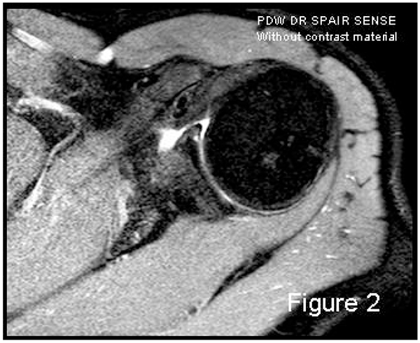
Figure 1. MRI image using joint effusion as contrast material in the acute setting.
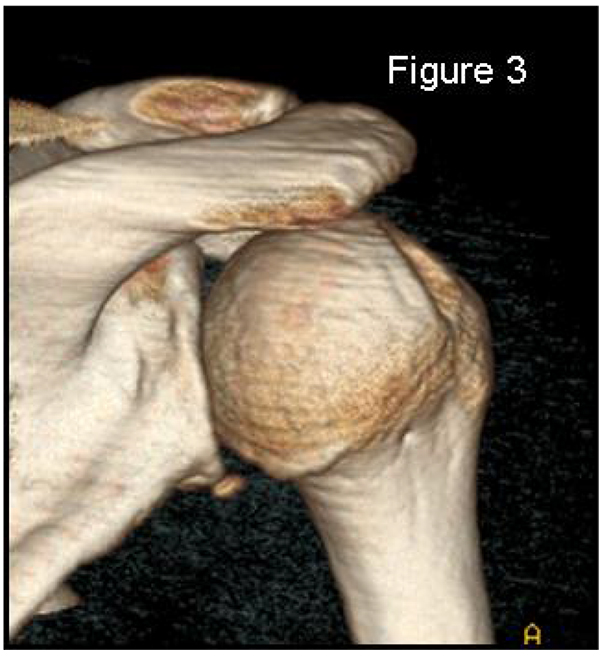
Figure 2. 3D CT is the gold standard for evaluating and quantifying the humeral head bone defect.
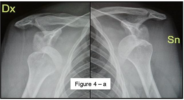
Figure 3. Bilateral Hill-Sachs lesions on an anteroposterior radiograph.
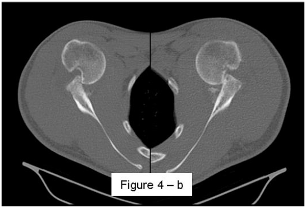
Figure 4. Axial CT showing bilateral engaging, wide and deep Hill-Sachs lesions.
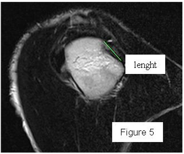
Figure 5. Measurement of Hill-Sachs lesion length on a sagittal CT view.
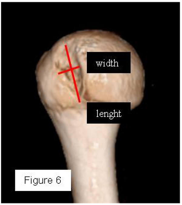
Figure 6. 3D CT en face view of the humerus with the scapula removed: the most accurate method to quantify the humeral bone defect (the minor axis is the width, the major axis the length).
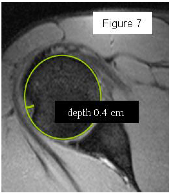
Figure 7. Measurement of humeral defect depth on axial CT using the circle method.
Figures from Baudi et al. [1], CC BY 4.0.
References
- Baudi P, Rebuzzi M, Matino G, Catani F. Imaging of the Unstable Shoulder. Open Orthop J. 2017;11:882-96. doi:10.2174/1874325001711010882.
- Applegate GR, Hewitt M, Snyder SJ, Watson E, Kwak S, Resnick D. Chronic labral tears: value of magnetic resonance arthrography in evaluating the glenoid labrum and labral-bicipital complex. Arthroscopy. 2004;20(9):959-63. doi:10.1016/S0749-8063(04)00858-8.
- Bushnell BD, Creighton RA, Herring MM. Bony instability of the shoulder. Arthroscopy. 2008;24(9):1061-73. doi:10.1016/j.arthro.2008.05.015.
- Kim DS, Yoon YS, Yi CH. Prevalence comparison of accompanying lesions between primary and recurrent anterior dislocation in the shoulder. Am J Sports Med. 2010;38(10):2071-6. doi:10.1177/0363546510371607.
Author: Olena Vyrva, MD
Questions & Answers
Q1. When is MR arthrography preferred over conventional MRI?
After an acute traumatic dislocation, the joint effusion or haemarthrosis can act as contrast on conventional MRI, so MRA is best reserved for chronic instability.
Q2. How much glenoid bone loss is considered critical?
There is no consensus, but most authors place the limit at about 20–25% of the glenoid surface; a 6–8 mm defect corresponds to 20–25% bone loss, the critical value for choosing bone augmentation.