The effect of various types of physical activity on the skeletal system in women
Relevance
Osteoporosis is a problem that may develop in women during the postmenopausal period. What could be so alarming about a decrease in bone mineral density (BMD)? However, as we know, such changes may have adverse consequences, including reduced social activity among women, increased risk of injury, and even a threat to life. According to the article, in 2011 more than 1.7 million people were hospitalized with fractures associated with increased bone fragility, resulting in 70 billion in direct treatment costs. The authors also present the following US statistics: women with osteoporosis older than 50 years have an increased risk of developing fractures during the subsequent decade compared with women with normal BMD (3.4% versus 6.8%). Looking at Ukrainian osteoporosis statistics, published data indicate that 13.4% of women and 2.9% of men in the country have this condition (Povorozniuk V.V. et al). Other sources report progressive growth of osteoporosis in Ukraine among 20-39% of the female population and 9-23% of men, depending on the region of residence. The risk of pathological fractures reaches 10%. Data from the International Osteoporosis Fundation indicate that 7 million women in Ukraine, approximately 28% of the total female population, have osteoporosis or osteopenia. We have all heard that diseases are much easier and less expensive to prevent than to treat. Osteoporosis in postmenopausal women illustrates this principle particularly well because this serious condition can be attenuated or even prevented through appropriate physical activity. Moreover, such activity should be started as early as possible. For example, the original article presents data suggesting that the onset of osteoporosis can be delayed by 13 years if bone mass is increased by 10% during the period of peak growth, which in turn may be achieved through appropriate nutrition and a suitable physical activity regimen. The effectiveness of exercise decreases later in life, but even at the age of 60 years and older, an appropriately selected exercise program may reduce the probability of a pathological fracture. In Ukraine, a group of researchers (Tyshchenko I.V. et al) conducted an interesting study examining women's awareness of osteoporosis and methods of addressing it. All 95 respondents had major risk factors for osteoporosis, including age over 65 years, smoking, early menopause, concomitant disease, and others. Most Ukrainian women in this cohort (more than 50%) considered it necessary to increase physical activity through regular exercise and to monitor their diet with an emphasis on calcium-containing foods.
Material and Methods
In this article, the authors collected what they considered the most illustrative examples of the demonstrated effects of exercise on women's bone tissue in different age groups, discussed the optimal timing for starting specific types of exercise and their duration, and addressed other related issues. The response of bone to mechanical influences and diagnostic criteria used to study the response of bone tissue to axial loading were also considered.
More relevant information is available via The biological clock: how ovarian aging impacts female fertility
Bone itself is a natural composite material that, according to the article, consists of 75% calcium salts, 23% type I collagen, and 2% ground substance, with all figures calculated on a dry-weight basis. As we know, bone also contains specifically arranged trabeculae that, similar to arches in engineering structures, allow maximum strength to be achieved with minimal mass. These characteristics enable bone tissue to withstand substantial loads. However, everything has its limit, and when a force exceeds the strength limit of a bone, the bone fractures. Obviously, it is impossible to measure the exact strength limit of a particular bone in a living person, but it can be calculated from the size, thickness, and structure of the bone and its volumetric bone mineral density (vBMD), expressed in g/cm3. With age, the cortical layer becomes thinner and bone vBMD decreases, which in turn reduces bone strength. Dual energy X-ray absorptiometry (DXA) is used to measure bone mineral density. This method makes it possible to assess the amount of the mineral component of the bone tissue under examination. Because the calculation is performed using a two-dimensional radiograph, the unit of measurement is g/cm2. BMD is calculated from the resulting value. These data are subsequently used to calculate the T-score and construct a scale of standard deviations in the population according to age and sex. On this scale, zero corresponds to the BMD value obtained from young, healthy individuals. As a reminder, the T-score is the difference between the BMD value of a specific individual and the mean BMD values in the population, taking sex and age into account. According to information in the article, the World Health Organization (WHO) defines osteoporosis as a T-score of –2.5 or lower. When combined with medical history data, the T-score may serve as a predictor of a possible fracture. Despite extensive experience with its use, DXA has important limitations related to its two-dimensional nature. For this reason, a three-dimensional method for measuring BMD based on computed tomography (CT) was developed. It allows a larger number of parameters to be calculated for assessment of bone condition, but involves greater radiation exposure and is less accessible than DXA (Fig. 1). In addition, three-dimensional BMD assessment makes it possible to investigate structural changes in bone tissue, which is important when evaluating the response to treatment.
Results and Discussion
Because bone tissue is living tissue, it has a certain capacity for adaptation and can change depending on the level of load placed on it, a process referred to as functional adaptation. In their work, the authors identify the following main indicators of bone strength: BMD, maximum cross-sectional area, and moment of inertia. According to studies, all these parameters are higher in people who participate in sports, particularly gymnastics. A similar difference is observed in tennis players when comparing the playing and non-playing arms. During physical activity, mechanical oscillations occur in bone, and their characteristics depend on the magnitude of the applied force, which may act externally through gravity and inertia or internally through contraction of the person's own musculature. Osteocytes detect these changes and initiate a response involving stimulation of osteoclasts for bone resorption and osteoblasts for enhancement of bone formation. These cell groups function in parallel, and their activity is continuously regulated according to the load applied to the bone. Because weaker bones develop substantially greater physiological movement-related deformation than stronger bones under comparable loads, the resulting remodeling response is also more pronounced. Taking all of the above into account, it is important to understand that exercise-induced changes in bone are complex and depend not only on the condition of the bone tissue but also on the characteristics of the load: bone undergoes greater remodeling in areas exposed to the highest load. Under normal conditions, bone remodeling occurs through coordinated activity of osteoblasts and osteoclasts. Osteoclasts are responsible for bone resorption, which is regulated by parathyroid hormone, but according to the authors it remains unclear what causes osteoclasts to resorb more bone tissue in one location than another. It is also unclear how these cells regulate the volume of resorption. There is evidence that influences on bone tissue, including loading, minor damage, and electromagnetic fields, affect sites of osteoclast activity. Osteoblasts largely “work” in the same locations as their antagonists. The authors emphasize that bone formation occurs at a rate of 1 μm/day, whereas resorption occurs at 40 μm/day. Thus, even a relatively small imbalance in the degree of osteoclast and osteoblast activation may lead to substantial bone loss, as occurs, for example, during the postmenopausal period, when reduced estrogen levels no longer exert an adequate inhibitory effect on osteoclasts, resulting in systemic diffuse osteoporosis. In addition to physical loading and hormone levels, the concentration of vitamin D in the blood also influences the adaptive response of bone tissue.
As noted earlier, bone tissue continuously undergoes remodeling, which depends on age, sex, the type of physical activity, and its duration. According to the authors, the principal factor is nevertheless the age period during which regular physical activity begins: prepubertal, early pubertal, adolescent, young adult, or mature age. According to the article, by the age of 16 years girls have accumulated 80-90% of their bone mass, with approximately 50% formed during the first four years after menarche. Peak bone mass is reached at approximately 18 years of age, after which no substantial changes occur for 30 years. Children's bone tissue is highly sensitive to loading, and appropriately selected physical exercise promotes bone growth and accumulation of bone mass. This effect subsequently becomes established and persists for a long period. According to the authors, the most advantageous exercises in this respect are those that produce large-amplitude bone deformation over a short period of time. One example is a study in which a 1-6% increase in bone mass was observed in adolescent girls after six months of jumping exercises. A distinctive feature of increasing bone mass and bone strength at this age is that the results persist for many years after exercise has stopped. It should be noted that bone mass also increases during adolescence and youth without exercise, but the increase is substantially smaller than in physically active peers. In adulthood, exercise no longer has such a pronounced and long-lasting effect on bone strength and shape. In people older than 60 years, no exercise increases bone mass, but exercise may prevent its decline. During the postmenopausal period, women lose an average of approximately 0.5% of bone mass per year; therefore, controlled physical activity is an important component of preventing postmenopausal osteoporosis. The premenopausal period also deserves attention because, according to the article, strength training can increase bone strength by 0.5% to 2.5%, thereby creating a reserve against possible bone loss after menopause.
The International Osteoporosis Fundation (IOF) recommends a range of exercises for preventing bone fragility that are referred to in English as weight-bearing exercises (WBE). In a free translation, this expression can be interpreted as “exercises with one's own body weight.” In English-language medical literature concerning osteoporosis, this term refers to physical activity in which a person works against gravity while remaining upright. WBE are divided into “high impact exercises,” including jumping rope, running, intensive aerobics, tennis, and dancing, and “lower impact exercises,” including walking, yoga, leisurely cycling, and use of elliptical and/or step machines. For convenience, we will refer to the first group as WBEH and the second as WBEL. According to the data in this review, the most effective exercises for people with osteoporosis are those in the WBEH group, which have a positive effect on bone condition at any age. Small studies have found that such loads significantly increase BMD in the vertebrae and femur in postmenopausal women. Resistance exercises, involving work with light weights and resistance bands while standing, also have a positive effect on bone strength during adolescence and adulthood. WBEL, which are commonly recommended for older people, do not produce the necessary effect on bone tissue. Yoga has only a minor effect on BMD in the spine, cycling affects the bones of the lower extremities, and swimming sometimes has a negative effect, causing a reduction in BMD. The authors recommend alternating all of these forms of activity with strength training to obtain the combined effects of different exercise groups. The article draws attention to the fact that although WBEH provide the greatest benefit to bone, they may adversely affect joint condition. Therefore, for people with confirmed osteoarthritis (OA), a gentler form of exercise is recommended. The authors associate this approach with the observation that loading of healthy articular cartilage causes it to thicken and improve, whereas similar loading of damaged cartilage leads to thinning and degeneration.
Running may improve cardiovascular condition and muscle strength, but it is often associated with a high risk of joint injury. At the same time, a meta-analysis of 17 studies involving more than 100000 people found OA in only 3.5% of recreational runners, whereas the figure reached 10.2% among people with a sedentary lifestyle. Notably, the highest percentage of OA occurred in professional runners, at 13.3%. These figures indicate the need for a rational approach to running and realistic assessment of one's capabilities. Otherwise, the expected health benefit may be replaced by deterioration in health. According to the authors, a similar situation applies to people who decide to use running as a means of improving cardiovascular health. In any case, before beginning any physical activity at an older age, a thorough consultation with a physician is necessary.
One of the major problems researchers encounter when investigating bone loading is the difficulty of obtaining accurate data showing the degree to which a particular physical exercise affects bone tissue. When working with animals, biological material can be collected. In human studies, this option is excluded, and several alternatives have therefore been proposed. In 1975, researchers placed strain-gauge sensors over bones to record changes occurring in the bone under different types of loading. A brief editorial note is appropriate here. A strain-gauge sensor is a special device that makes it possible to convert the degree of deformation of a structure exposed to a specific force into measurable numerical values. The most common household device of this type is a weighing scale. As can be expected, this method has two important limitations. First, measurements are possible only in locations with a small thickness of soft tissue. Second, because the devices are relatively bulky, long-term placement is extremely difficult. Subsequently, a special technique was developed that made it possible to measure deformation of the tibia during loading using special screws inserted into the periosteal layer. Precise measurement of displacement of the screw heads provided accurate information about loads on the bone. This technique is accurate but, as correctly noted in the article, is not applicable in clinical research. In their work, the authors decided to use a combination of methods: sensors measuring force and calculations based on the finite element model (FEM). FEM is a special method in applied physics that uses solutions to integral and differential equations to study the mechanics of deformable solid bodies. In this way, the degree of bone deformation can be calculated according to the loads applied to it. Using volunteers, the authors studied deformation of upper-extremity bones occurring during palm support. A specified level of pressure had to be achieved. In addition to measuring the applied force, CT-based FEM was used to calculate deformation of the radius, lunate, and scaphoid bones. The authors found that in premenopausal women with normal BMD, identical loads produced varying deformations, which consequently affected bone remodeling differently. In our opinion, these findings demonstrated a high degree of individual variability in the human body, which in this case depended on the anatomical characteristics of the participant's upper extremities. Taking the above into consideration, the authors express the hope that specialists working in this field will in the future develop methods for measuring bone loading that account as fully as possible for all identified anatomical nuances.
Despite the availability of methods for measuring BMD, including DXA and quantitative computed tomography (QCT), researchers continue to propose new methods intended to improve measurement quality. These developments include 3D imaging and high-resolution QCT. The first method makes it possible to use QCT images to construct three-dimensional models and obtain a substantially larger amount of information for analysis (Fig. 1). There are two major barriers to widespread introduction of 3D imaging into clinical practice: high radiation exposure and high cost. The second method makes it possible to assess bone microstructure down to individual trabeculae. At present, the areas available for investigation are limited to the distal radius and tibia with first-generation devices and the knee joint with second-generation devices. When analyzing these areas, the authors found that in postmenopausal women individual trabeculae in the radius become thicker while their number decreases, and cortical porosity increases in the distal radius and tibia. Such analysis provides a better understanding of bone-loss processes in postmenopausal women and allows more precise assessment of the effects of specific physical exercises. In addition, QCT-based FEM was used by the authors to assess changes in a nine-millimeter layer of bone subjected to compression simulating mechanical loading. The aim of this study was to identify exercises that maximally increase bone strength and determine the exact load values that produce the required osteogenic response. However, during the study it became apparent that the modeling did not provide the required result because the mechanism by which the simulated load affected bone differed substantially from physiological loading. Therefore, the authors recommend combining different imaging techniques with multiple assessment scales for a more comprehensive evaluation.
Determining the response to short-term osteoporosis treatment may help not only to adjust subsequent therapy but also to optimize the necessary physical activity. For objective assessment, the authors recommend using several markers measured in urine and blood serum. Despite the large number of different tests for various markers, the International Osteoporosis Fundation (IOF) proposes using only two: measurement of propeptide and C-terminal telopeptide of type II collagen in serum, which serve as useful tools for monitoring bone resorption and bone formation. In addition to these parameters, the authors draw attention to sclerostin, changes in the level of which have been studied as a response to mechanical loading of bone. As an endogenous inhibitor of the Wnt/β signaling pathway, sclerostin suppresses bone formation by reducing osteoblast activity and increasing osteoclast activity. According to information in the article, animal studies have demonstrated an association between mechanical loading and blood sclerostin levels. Thus, the authors consider that measurement of sclerostin levels may become one of the objective criteria for evaluating the effectiveness of selected physical loads when studying the effects of exercise on bone tissue.
Because osteoporosis treatment is based on modifying bone remodeling to enhance osteoblast activity and reduce the functional activity of osteoclasts, it was suggested that accompanying pharmacological therapy with physical activity could improve treatment effectiveness. The authors present the results of seven randomized studies comparing two groups of patients. The first group was treated with hormonal therapy involving estrogen-containing medications and antiresorptive therapy with alendronate or risedronate in combination with physical activity, whereas the second group received medications alone. The first group showed a significantly greater increase in BMD in the lumbar vertebrae than the second group. This effect was not observed in the distal femur, which was attributed to the specific characteristics of the exercise program.
Additional relevant facts are available via Hormone therapy: friend or foe? Navigating the benefits and risks for menopausal women
Conclusions
Physical activity is one of the most important components of healthy bone. Based on the studies reviewed, the authors provide several recommendations intended to help structure appropriate physical activity:
- During the prepubertal and adolescent periods, exercises producing a high level of bone loading are recommended. Such practice may not only maximize the increase in bone mass but may also prevent fractures in the future.
- Among exercises producing high bone loading, the most effective, according to the authors, are jumping and hopping combined with resistance exercises.
- The duration of each exercise session should be approximately half an hour. According to the data obtained, sessions lasting less than 30 minutes produce changes only in the femoral neck. If improvement in spinal BMD is required, each session should last more than 30 minutes. Frequency should range from 2 to 4 times per week depending on the risk group of the individual exercising. The regularity of such activity must also be maintained. In addition, according to the research data presented in the article, the duration of training should be at least 16 years.
- For women in older age groups, exercises from the WBEL group are recommended. Walking is reported to have a positive effect only on BMD in the femoral neck, and even this effect is not always observed. Nevertheless, walking remains relevant for improving balance, thereby reducing the probability of falling and consequently the probability of fracture.
We have presented in this review what we consider the most interesting information included in the original article. To obtain a complete picture of the subject, we recommend following the link provided at the beginning of our review.
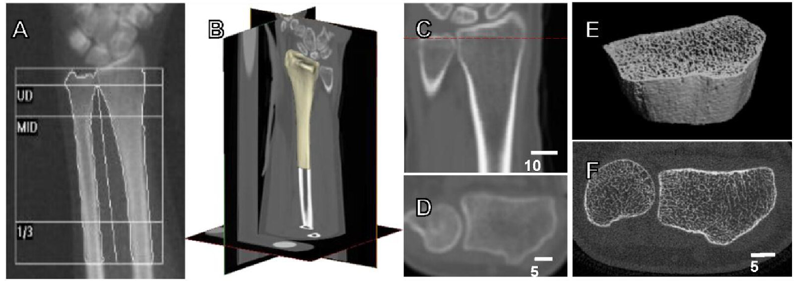
Figure 1. Methods for measuring bone strength and fracture risk. (A) Dual energy X-ray absorptiometry (DXA) of the forearm with BMD measurement. (B) 3D image of a CT scan of the distal radius with (C) a coronal view in which the level of the cross-section is marked by a red dashed line, followed by formation of the CT image (D). (E) High-resolution 3D CT image of the distal third of the radius (F).
References
Troy KL, Mancuso ME, Butler TA, Johnson JE. Exercise Early and Often: Effects of Physical Activity and Exercise on Women's Bone Health. Int J Environ Res Public Health. 2018;15(5):878. Published 2018 Apr 28. doi:10.3390/ijerph15050878
Author: Olena Vyrva, MD
Questions & Answers
Q1. At what stage of life does physical activity appear to have the greatest influence on bone mass according to the review?
The article emphasizes the prepubertal and adolescent periods. By age 16, girls have accumulated 80-90% of their bone mass, and exercise-induced gains during this period may persist for many years.
Q2. Which types of exercise were described as having the greatest positive effect on bone?
The review identifies high-impact weight-bearing exercises, particularly jumping and hopping, together with resistance training as the most effective approaches for increasing or preserving bone strength. Their suitability may vary with age, joint condition, and individual risk.